This article just goes to show how adaptable the human body is. The featured woman was 24 years old when it was discovered she is missing her whole cerebellum and despite a few struggles with motor skills and speech, she manages just fine. It begs the question: could we survive with other parts of our brain missing? The article says it is not unheard of to be missing parts of the brain. We know that a missing cerebellum creates a few impediments, but what does a missing brain part mean for longevity, or long-term health concerns?
From my research, it appears that theoretically a person could survive without a parieto-occipital sulcus. The parieto-occipital sulcus is the ridge between the parietal lobe and the occipital lobe. person may also have a connected parietal and occipital lobe, leading to a whole new breed of problems. It appears to help with planning things - so maybe a person would struggle with forethought without it. The frontal lobe is also involved in planning, so it is possible that if the parieto-occipital sulcus was missing, the frontal lobe could just take over.
Wednesday, April 13, 2016

Thursday, March 24, 2016
Unit 7 Reflection
Unit 7 is all about the muscular system. We began with how synovial joints let us perform different actions, for example, flexion and extension. We created mini dances that included all the different movements and performed them in front of the class. It was all in good fun, but also uniquely helpful in memorizing each different synovial movement. We also learnt the basic anatomy of muscles: the major muscles in our body, and in a chicken (by dissection), such as the major and minor pectoralis, the deltoid, and the trapezius. The dissection was really cool - seeing all the different muscles and how similar they are to a humans was really impressive.


We then moved onto how muscles are separated and classified into different groups. Next, we zoned in and learnt about how muscles specifically work, right in each muscle fiber. I found the sliding filament theory particularly interesting - how the muscle shortens and lengthens when contracting and relaxing. We made a short video showing how a muscle works. It was challenging, finding a way to portray it clearly, but it was a great learning experience and it was cool how we made an actual short movie on it. Next, we moved onto how muscle fibers respond to different exercise - slow oxidative fibers in marathoners, fast oxidative fibers in a regular person, and fast glycolytic fibers in high intensity/ short burst workout, like sprinters. Finally, we covered performance enhancing substances and how the affect the mind body. The mini project for this section was that we made a satirical ad promoting P.E. substances.
 The thing that really stood out to me about this is that none of them are FDA approved, or regulated in anyway, which in turn makes them all very unsafe for consumption.

I'm really interested in learning more about performance enhancing substances, not only in the body, but those made for the mind as well. They are all so dangerous but many people treat them like no big deal - maybe not the ones aimed at body-building, but the ones that aid in focus and improve mental performance. Overall, I like a lot how my 20 time project is coming along, I'm really satisfied with my progress so far. Going back to my New Year's Goals, I am still working on more actively participating in class but in small groups I am participating a lot more. I am sleeping a fair amount every night - losing an hour to daylight saving wasn't great but I'm making it work the best I can. For horse riding, I am working hard to get to where I want to be - and hopefully a new horse is coming my way in the near future!
Wednesday, March 23, 2016
Performance Enhancement Advertisement

Thursday, March 17, 2016
Chicken Dissection Analysis
We skinned the chicken and got a good look at all the muscles, first and foremost the pectoralis major and minor. The muscles help the chicken move and hold itself up. The allow for the chicken to walk, hop, stand, and sit. For example, the pectoralis major gives chickens the ability to move its wings ventrally. The pectoralis minor allows the chicken to move its wings dorsally. The bones give the chicken its skeleton and are what allows it to have the anatomy to move. The tendons attach the muscles to the bone so that the muscles can move in different directions, which in turn allow the chicken to perform movements.

The insertion of the muscle in the wings are quite shiny and white. When they move, the muscle is lengthened or shortened, and they glide against each other - as the chicken would when performing that movement.
Surprisingly, the chicken's muscles were quite similar to those of a human. The chicken has biceps and triceps like humans, obviously on a much smaller scale, but we quickly identified them. The trapezius was on the back, same as in humans, despite the overall difference in anatomy. It was also not hard to identify. The sartorius are on the front of the thigh, and allows for the crossing of the legs. Same on a human, they were also pretty easily spotted. The shape of a chicken is very different from a humans, but the muscles it has are not so different.
Surprisingly, the chicken's muscles were quite similar to those of a human. The chicken has biceps and triceps like humans, obviously on a much smaller scale, but we quickly identified them. The trapezius was on the back, same as in humans, despite the overall difference in anatomy. It was also not hard to identify. The sartorius are on the front of the thigh, and allows for the crossing of the legs. Same on a human, they were also pretty easily spotted. The shape of a chicken is very different from a humans, but the muscles it has are not so different.
 |
| Pulls the hand back |
 |
| Flexion of the leg |

 |
| Includes sartorius, iliotibialis, biceps femoris, semimebranosus, semientendinosus, and quadriceps |
 |
| Extends foot, flexes lower leg. Allows us to stand on our toes |
 |
| Flexion of the hand |
 |
| Extends the thigh, flexes the leg |
 |
| Flexes the thigh, extends the lower leg |
 |
| Deltoid: raise the upper arm/ wing Biceps: flexion of arm/ wing Triceps: extension of arm/ wing |
 |
| Pulls the wing ventrally, in most birds it allows for flight |

 |
| Lifts the wing dorsally, or in humans in pulls the shoulder down and forward |
 |
| Extends the thigh |
 |
| Pull the shoulder back |
 |
| Extends the thigh |
 |
| The white, shiny tendon in the wing that attaches the bone to muscle |
 |
| Flexes the thigh, allows crossing of legs |
 |
| Our chicken, before any cuts were made |
Monday, March 14, 2016
What Happens When You Stretch?
1) "Hence when you stretch, the muscle fiber is pulled out to its full length sarcomere by sarcomere, and then the connective tissue takes up the remaining slack."
- This quote explains what happens when stretching -- how the muscle lengthens fully to accommodate what is being asked of the body. There are many sarcomeres, and a full stretch occurs when all of the sarcomeres are fully extended.
2) "Some sources suggest that with extensive training, the stretch reflex of certain muscles can be controlled so that their is little or no reflex contraction in response to a sudden stretch."
- Some think that if people train enough, they can control which muscles stretch and which ones contract. The less contraction, the more stretch possible. It is not easy to achieve; as the sources say only the top of the top athletes achieve it. -- but it is very rewarding once achieved.
3) "When stretching, it is easier to stretch a muscle that is relaxed than to stretch a muscle that is contracting. By taking advantage of the situations when reciprocal inhibition does occur, you can get a more effective stretch..."
- To increase the ease of stretching, it is very important to be relaxed , so contracting muscles are not opposing. Contracting certain muscles while trying to stretch others is very contradictory when trying to elongate the muscle.
Relate & Review
This reading was about what happens when you stretch. It's important to try to control the muscle contraction when trying to stretch =, so the sarcomeres can fully extend and stretch out as much as possible; and also to improve the ease of stretching muscles. Gymnasts and dancers seem to be about to control when the muscle contractions occur, which totally changes the game when the sports are both very focused on extension of limbs. As a former gymnast and dancer, it is easy to recognize how helpful it is the be able to control sudden muscle contractions.
Monday, February 29, 2016
Unit 6 Reflection
This unit was about the skeletal system: the structures of bones, disorders of the skeletal system, bone remodeling, bone fractures and how they are repaired, the bones in the body, and joints. Straight off the bat, it was very interesting to see that bones are composed differently which is why they are separated into types. I also did not know that bones are constantly breaking down (osteoclasts) and reforming (osteoblasts). We learnt how the skeleton is separated up and what the bones do, and how they completely affect our daily lives, from running to even just sitting up right. The disorders of the skeletal system were interesting to learn about - my closest friend has had scoliosis since she was 10 years old, but swimming has helped with it. The video we watched in class about correct posture and how it affects us made me really think about how often I slouch and the weight my neck has to bear from constantly looking down at my phone. Breaking bones is not necessarily common, but a lot of people I know have broken at least one bone in their body. Seeing how the bone will repair itself, and then be as good as new, was fascinating. I never really noticed how important joints are in the body but they have a direct impact on bodily movement, so are very necessary. I would like to learn more about the necessity of joints for bones. The owl pellet lab helped me understand the skeleton a lot better, and how similar an animal skeleton is to a human skeleton. Our owl pellet skeleton was very incomplete, but it goes to show how few bones are necessary to identify what organism it was. I found this unit to be very interesting, I previously knew very little about bones. Now I see an x-ray and am interested in figuring out which bone is which. My New Year's goals are going well, I am constantly growing in class and becoming more comfortable around my classmates. I am getting ahead in my horse riding and am setting up with a horse that will help me accomplish my goals. I have been sleeping quite regular hours, but I could definitely sleep more. I'm also excited to see where my 20 times goes, I really like my topic and I am excited to see where I can take it.

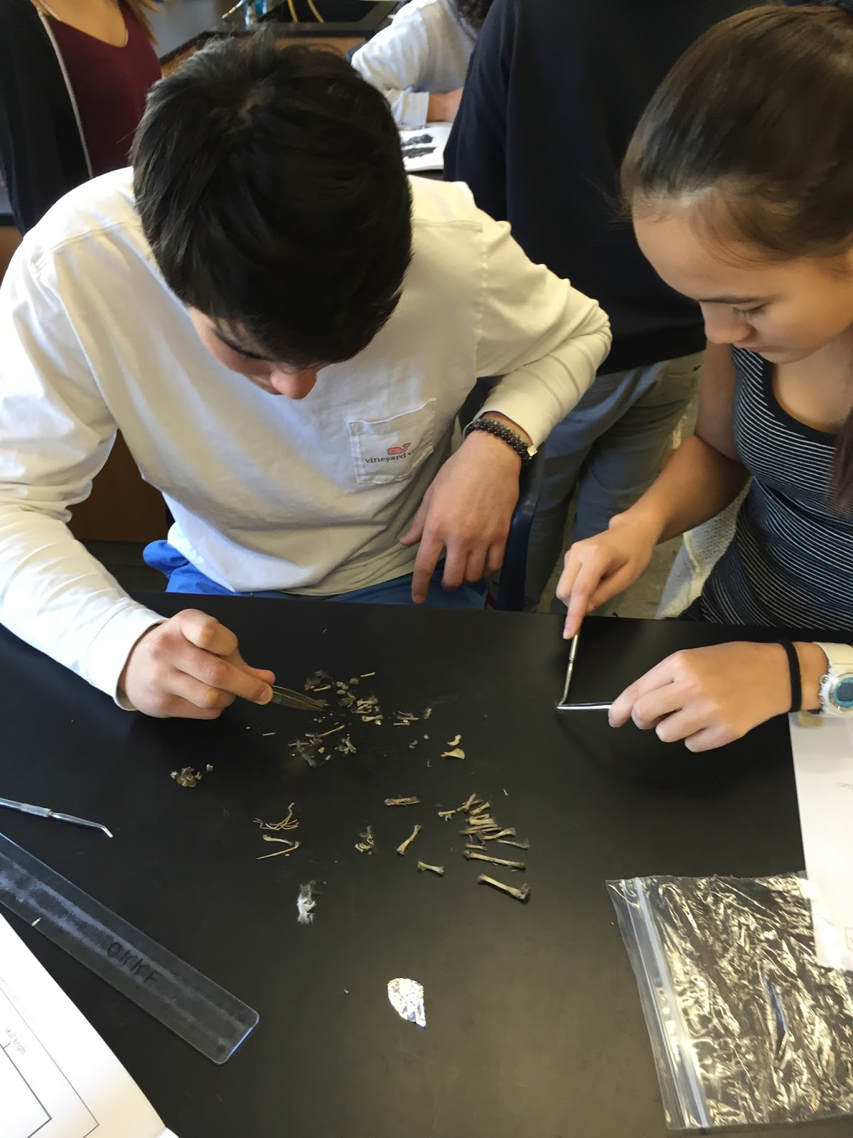

Thursday, February 25, 2016
Owl Pellet Lab
In the owl pellet lab, we were given an owl pellet (indigestible remains owls regurgitate), and broke it apart in an attempt to find out what the owl had eaten. We used forceps and a probe to pull apart the compact mass and found a lot of fur, and a broken up skeleton. We separated the bones into groups based on similarity, and then tried to find out what it was.
Our owl pellet skeleton was quite incomplete, there was no complete skull, which made it difficult to distinguish what it was. However, we decided that the organism was a vole -- based solely on the shape of the scapula, and the radius and ulna. The diagram of the scapula for a vole was the only one that matched up with the physical scapula bone of our organism. The radius and ulna did not match at the ends of the bones for the other small rodents, and the diagram for the radius and ulna matched the organism's bone the most closely. Despite the incomplete skeleton, we strongly concluded that it was a vole.
Our owl pellet skeleton was quite incomplete, there was no complete skull, which made it difficult to distinguish what it was. However, we decided that the organism was a vole -- based solely on the shape of the scapula, and the radius and ulna. The diagram of the scapula for a vole was the only one that matched up with the physical scapula bone of our organism. The radius and ulna did not match at the ends of the bones for the other small rodents, and the diagram for the radius and ulna matched the organism's bone the most closely. Despite the incomplete skeleton, we strongly concluded that it was a vole.
The scapula and ulna and radius looked very similar to human bones -- not at all in size, but the shape of them could almost have been the same, just shrunken. In the scapula, there was a center ridge that humans do not have, but it was easy to tell that it was the same bone, just in a vole. The spine was all separated into different sections, but we also distinguished very quickly that it was the spine. It looked just like a human skeleton, of course again on a much smaller scale. A human radius and ulna and a vole radius and ulna look incredibly similar, again just minimized.
 |
| Vole and shrew leg bone comparison charts |
 |
| Spine |
 |
| Close up of scapula compared to diagram |
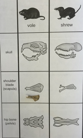 |
| Vole and shrew comparison chart (skull, scapula, pelvis) |
 |
| Close up of ulna and radius compared to diagram |
Subscribe to:
Posts (Atom)